Top Banner For Antibiotics

Top Banner For Antibiotics
get the full information...

вторник, 30 ноября 2010 г.

Multiple use of antibiotics


In the feature article “Intracellular Activity, Potential Clinical
Uses of Antibiotics” Robert M. Rakita (ASM News, 64, 570, 1998)
discusses the three-way interaction of the pathogens, host defense
cells, and antimicrobial agents, especially inside the neutrophils
and macrophages. The preparation of newer macrolide and
quinoline antibiotics try to achieve higher intracellular levels with
greater antimicrobial activities and with minimal cellular damage.
The goal being the control and elimination of the infecting bacteria.
What is often over-looked is the broad cellular reactions of
antibiotics in addition to their antimicrobial and clinical response.
Inhibiting a pathogen’s growth with antibiotics usually includes
the inhibition of cellular protein synthesis while its elimination
depends on its intracellular location and the host’s immune
responses. Many more conditions can effect the reactivity of
pathogens and antibiotics in the complex host tissues than in the
controlled in-vitro Tissue Cell Cultures. The variable tissue
pathogenicity also contributes to the variable antibiotic sensitivity
requiring adjustments for each pathogen and their tissue
location.(1)
Prior to the availability and application of antibiotics for the
control of diseases, gold salts, arsenicals, sulfa drugs and other
various chemicals were used to ward off the offending bacterial
pathogens without killing the patient. The clinical application of
antibiotics started in the early ‘40’s when penicillin became
available during WWII as the first miracle drug from a penicillium
mold. A few years later when the broad spectrum tetracycline
antibiotics became available their clinical use spread like a gold
rush. The bacterial sensitivities and potential clinical application of
the new antibiotics were extensively used by clinics and
laboratories. The choice of antibiotics was made after the isolation
and identification of the infecting agent. The development of
allergies and toxicities to some antibiotics limited their use in some
patients. Microcidal penicillins inhibit bacterial cell wall synthesis,
whereas the tetracyclines (macrolides) are micro static inhibiting
protein synthesis and the growth of the wall-less bacteria such as
mycoplasmas.
The initial use of high dosage antibiotics in some chronic
disease patients may cause a flare or clinical worsening with a
serologic rise in antibody titer to a suspected microbial agent such
as mycoplasmas. A temporary flare of symptoms following
antibiotic treatment is often referred to as a Jarisch Herxheimer
reaction. The flares often occur in joints or areas that have been
quiet or dormant since the arthritis was first observed. Knowing
this the patients are encouraged by the temporary worsening
following their antibiotic treatment. The delayed reaction resulting
from the release of microbial antigen into the sensitized host tissue
as in a “Graft vs. Host” reaction that is not a drug sensitivity.
Similarly the occurrence of physical &/or mental stress could also
initiate clinical worsening with a rise in microbial antibody titer.
The flare reaction could also result from the released microbial
antigen complexing with its circulating antibodies to promote
Complement Fixation. The antibiotics, tetracyclines, can also act
like the immunosuppressant steroids by blocking the formation of
the antibiotic+antigen complex that initiates inflammation. Many
clinical disorders are considered Immune Complex Diseases of
infectious origin, such as rheumatoid arthritis and Lupus, resulting
from the activation of complement and proteolytic destruction of
tissues with the deposition of Immuine Complex on the kidneys
and other tissue cell membranes.
The tetracycline antibiotics are potent metal chelating,
complexing, agents and comparable in action to the clinical use of
the chelating agents ethylenediaminotetraacetate, EDTA, and
penicillamine. Consequently the mode of antibiotic administration,
Intravenous or Oral (between meals), could have an affect on
the composition of their absorption state and thus their reactivity.
When complexed with divalent trace metals (Cu, Zn, Mg, Se, etc.)
The antibiotics become antioxidants or electron scavengers. As
such the metal antibiotic complex becomes antiinflammatory
neutralizing free oxygen radicals. By combining with metaloproteins
and metaloenzymes such as collagenase, antibiotic therapy can
inhibit collagen tissue destruction. If used excessively in high doses
the antibiotics, as protein synthesis inhibitors, could also inhibit the
synthesis and function of essential cellular proteins and not just the
pathogens.
Because of their immunosuppresive actions the macrolide
antibiotics can block and limit the immune complex (Antibody +
Antigen) formation and thus stop the complex induced
inflammation. In cases with low pathogenic activity, such as
mycoplasmas, pulsed antibiotic therapy with lower doses over
longer periods has proven more effective and with fewer side
effects. Tissue cells will survive intermittent (pulse) treatment of
tetracyclines but not constant exposure even at lower doses. In the
chronic immunologic disorders of probable infectious etiology
high daily antibiotic doses are not essential or effective for the less
virulent agents.
Bioassays for antibiotic levels in blood and tissues measures
the antimicrobial action that would not explain their other activities
based on intracellular concentration.
Although suspected of infectious origin the clinical trials of
minocycline antibiotic in rheumatoid arthritis was based primarily
®
Medical data is for informational purposes only. You should always consult your family physician, or one of our referral physicians prior to treatment.
on its inhibitory action of the metalloenzyme, collagenase, that
destroys the connective tissues and joint cartilage causing the
inflamed joints.
The effectiveness of treatment with minocin antibiotic was
based primarily on the eradication of arthritis inflammation rather
than infectious agents. The maximum effectiveness of the
antibiotic treatment was found in the duration of therapy indicating
a slow healing process that has to balance cell growth versus
inhibition of protein synthesis and microbial growth by the
multiple antibiotic actions. Growth inhibiting antibiotics may
control mycoplasma or microbial growth for an indefinite period
until the neutralizing antibodies and immune system process their
elimination.
Antibiotics can be used in the identification of the infectious
bacterial agent(s). In cases where the agent can not be isolated and
identified or the DNA can not be matched it is possible that
antibiotic therapy will cause the release of the microbial antigen to
initiate a specific antibody response. The serologic measure of a
change or response in the serum antibody level to a bacterial
infection would indicate its presence. The sero conversion or the
increase in antibody titer, resulting from the administration of a
vaccine would indicate the host’s immune responsiveness to a
particular antibiotic therapy. The specificity and sensitivity of the
serologic response depends on the test used, such as: growth
inhibition, neutralization, agglutination (ELIZA), complement
fixation, immunoblotting.
A rise in serum antibody level during the acute to convalescent
phase on antibiotic therapy would indicate a concurrent infection or
the antigen release from a persisting silent infection. A similar
positive sero conversion with a rise in antibodies could be observed
in a patient following physical or mental stress. No rise in antibody
titer to a vaccine, infection or stress would indicate an
immunodeficient agammaglobulinemia subject with limited
antibody production and immune defence. In rheumatoid arthritis
and other infectious diseases that initiate the anti-antibody response
(rheumatoid factor) RF the antibody levels are inversely related
causing an apparent decrease or negative sero conversion.
Following antibiotic treatment when the mycoplasma antibody
level increases, the RF test results will be lower.
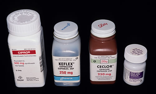
The use of generic antibiotics may have the same
antimicrobial potency while their systemic action in the host may
varay significantly. For example in the treatment of RA the generic
minocycline is reportedly less effective than minocin. In some
patients this difference in antibiotic action could result from patient
differences.

суббота, 20 ноября 2010 г.

Modification of the antibiotic olivomycin

The aureolic acid family of antitumor antibiotics includes a group of
clinically active agents such as olivomycin I (olivomycin A), mithramycin,
and also chromomycin A3 and durhamycin.1 The antibiotics of
the aureolic acid family interact with the DNA minor groove in high-
GC-content regions in a nonintercalative way and with the requirement
for Mg2+ ions.

The antitumor antibiotic olivomycin I was discovered at the Gause
Institute of New Antibiotics, Moscow.3 Comparative study of the
antitumor action of olivomycin I and chromomycin A3 in in vivo
experiments on murine lymphosarcoma LY01 revealed that the chemotherapeutic
index (LD50/DIT50) of olivomycin I is more favorable
(2.35) than that of chromomycin A3 (0.99). A similar study on the
inhibitory effect of olivomycin I, chromomycin A3 and mithramycin
against transplantable murine leukemia La showed that a similar
antitumor effect (an increase in the lifespan of mice by 25%) can be
achieved at lower doses of olivomycin I than those for the other
aureolic acid antibiotics studied. Clinical investigations of mithramycin
and olivomycin I showed that these antibiotics give favorable results
in treatment of testicular tumors. It was shown that these antibiotics
exhibit side effects such as gastrointestinal, hepatic, renal and bone
marrow toxicities. The major clinically limiting toxicity of mithramycin
was a hemorrhagic diathesis associated with a precipitous thrombocytopenia.
It is of considerable interest that hemorrhagic diathesis
was not observed after administration of olivomycin I.

As olivomycin I possesses the best chemotherapeutic index among
the aureolic acid antibiotics, it can be considered as the best scaffold
for the development of novel semisynthetic aureolic acid analogs with
increased therapeutic indices and lower toxicity compared with the
parent antibiotic.
Here we describe chemical modifications of olivomycin I at the
2ў-keto group of the side chain of the aglycone moiety. The
antiproliferative and topoisomerase I (Topo-I)-poisoning activities
of the novel derivatives (2–7) were tested. One of the novel derivatives
showed pronounced antitumor activity in in vivo experiments on mice
bearing lymphocyte leukemia P-388, together with lower toxicity to
animals compared with the parent olivomycin I.

RESULTS

Chemistry
We developed a novel method of chemical modification of olivomycin
I (1) based on the introduction of a carboxyl group into the molecule
of the antibiotic. Reaction of olivomycin I (1) with carboxymethoxylamine
gave the key intermediate, 2ў-carboxymethoxime-olivomycin
I (2) (Scheme 1), which was further reacted with different amines in
the presence of PyBOP to give the corresponding amides 3–7. The
resulting compounds were purified by column chromatography on
silica gel.


Biological testing

The cytotoxicity of the compounds in comparison with the parent
olivomycin I was tested. Cells were incubated with drugs for 48–72 h
to ensure the completion of late events in cell death. Table 1 shows
the comparative potencies of these compounds against the
wild-type murine leukemia L1210 cells, the human leukemia cell
line K562 and the human malignant T-lymphocyte Molt4/C8 and
CEM cells.
All novel derivatives (2–7) caused cell death at higher concentrations
than olivomycin I. Remarkably, amides with the bulky
hydrophobic substituents (adamantyl- 5; tert-butyl-, 6) showed antiproliferative
activity that was at an IC50 of only one order of
magnitude higher (for L1210: 0.19 and 0.20 mM, correspondingly)
than that of olivomycin I, but at a markedly lower IC50 than that of
2ў-carboxymethoxime-olivomycin I (2) or the amides with small or
hydrophilic substituents 3, 4 and 7 (IC50 for L1210: 6.5–20 mM).
To identify tentative intracellular targets important for cytotoxicity
of olivomycin I and its novel derivatives (2–7), we tested these
compounds for their ability to modulate Topo-I activity in vitro.
Olivomycin I (1) and all novel derivatives (2–7) were potent Topo-I
inhibitors at all concentrations investigated (0.5–20 mM) (data not
shown). Figure 1 shows the results of electrophoretic analysis of the
relaxation products of the Topo-I-dependent supercoiled DNA relaxation
in the absence and presence of the antibiotics olivomycin I and 5.
In the absence of antibiotics (track of Topo-I), the reaction led to a set
of topoisomers and the disappearance of the supercoiled form of
DNA. This effect was revealed by the presence of residual amounts of
rapidly migrating topoisomers. Olivomycin I (1) inhibited Topo-I
activity at all concentrations investigated (0.5–20 mM); compound 5
was less active but still a potent Topo-I inhibitor—the rapidly
migrating topoisomers were observed on the track starting at the
compound concentration of 2.5 mM.

When 5 was i.p. injected to mice with leukemia P-388 72 h after i.p.
implantation of tumor, 62% increase of lifespan (ILS) at the dose
of 50mg kg was achieved.

Compound 5 did not show any cumulative toxic effects at the
quintuple doses of 5 and 10mg kg (25 and 50mg kg 1 total dose,
respectively), whereas all of the mice that received 1 (5 mg kg 1 daily)
had drug-related toxic death after the third injection (data no shown).
All mice that received daily doses of 5 and 10mg kg 1 had tumorrelated
death. The ILS was 43% compared with the group of untreated
mice (Figure 2). Quintuple injections of 5 at 20mg kg 1 per dose
(100mg kg total dose) resulted in drug-related death of 15% of the
mice.

Micafungin: a sulfated echinocandin

Fungal infections cause not only superficial diseases such as athlete’s
foot and onychomycoses, but also life-threatening diseases. Serious
deep-seated fungal infections caused by Candida spp., Aspergillus spp.
and Cryptococcus neoformans are a threat to human health. Incidences
of these systemic fungal infections have increased significantly over the
past few years. The major reasons for this dramatic increase are the
extensive use of broad-spectrum antibiotics and the growing number
of immunocompromised patients with acquired immunodeficiency
syndrome (AIDS), cancer and transplants.

In the mid 1900s, few compounds, such as polyenes (for example,
nystatin and amphotericin B) and flucytosine, were available for
antifungal chemotherapy. Although the development of azole drugs
started in the early 1970s, only a limited number of antifungal agents
were available for treatment of life-threatening fungal infections.
Moreover, the existing agents had disadvantages, such as the significant
nephrotoxicity of amphotericin B3 and the emergence of resistance
to the azoles.4 To overcome these defects, lipid formulations of
polyenes were developed to reduce toxicity, and new triazoles (for
example, voriconazole, ravuconazole and posaconazole) were developed
to improve the antifungal spectra or susceptibility to azoleresistant
isolates.5 Despite a number of therapeutic advancements,
there was a need to develop a new class of antifungal agents with novel
mechanisms of action.

The echinocandins were a new class of antifungal drugs developed
for the first time since azoles. The first launched echinocandin was
caspofungin acetate (Merck & Co. Inc. (Merck), Readington, NJ, USA),
followed by micafungin (Fujisawa Pharmaceutical Co., Ltd, now
Astellas Pharma Co., Ltd, Fujisawa, Japan) and anidulafungin (Vicuron
Pharmaceuticals Inc., now Pfizer Inc., New York, NY, USA), which was
originally developed by Eli Lilly and Company (Indianapolis, IN, USA)
(Lilly) as LY 303366 and subsequently licensed to Vicuron (formerly
Versicor) as VER-002. The approved echinocandins are synthetically
modified lipopeptides that originate from natural compounds produced
by filamentous fungi. The original anidulafungin, caspofungin
and micafungin compounds were echinocandin B from Aspergillus
nidulans var. echinulatus,6 pneumocandin B0 from Glarea lozoyensis7
and FR901379 from Coleophoma empetri,8 respectively.
Although natural echinocandins have potent antifungal activity
in vitro, their structures were chemically altered to improve their
absorption, distribution, metabolism and excretion characteristics.
Such operations were initiated by Lilly on echinocandin B to yield
cilofungin.9 This compound was subjected to Phase II clinical trials,
but was abandoned due to toxicity. Further modification of the
structure by converting the phenolic hydroxyl to a sodium phosphate
ester produced the more soluble prodrug LY307853, which resulted in
the active form, LY303366.10 Merck has produced MK-0991 using
pneumocandin B0 as the starting material.11 MK-0991 likewise possesses
increased water solubility. Other reviews on echinocandins or
individual antifungal agents have reported their usefulness in clinical
practice.12–16 This review describes the discovery and development of
micafungin, focusing on the chemical diversity of echinocandins.

DISCOVERY OF FR901379

The seed compounds of micafungin, FR901379 and two related
compounds (FR901381 and FR901382), were discovered at Fujisawa
Pharmaceutical Co., Ltd in 1989 from the screening of approximately
6000 microbial broth samples (Figure 1). These new compounds were
categorised as members of the echinocandin class of lipopeptides.
Echinocandin B, pneumocandin B0 and other echinocandin lipopeptides
are structurally characterized by a cyclic hexapeptide acylated
with a long side chain, and have an excellent anti-Candida activity
attributed to selective inhibition of 1,3-b-glucan synthesis, although
their intrinsic water insolubility is a major barrier for drug development.
17–19 However, FR901379 and related compounds showed both
high water solubility and a strong antifungal effect on Candida spp.20
The structural difference between FR901379 and the other echinocandins
is that FR901379 has a sulfate moiety in its molecule (Figure 1,
circled). This residue was speculated to be the basis for the high water
solubility of FR901379 (soluble in water even at a concentration of
50mgml 1, a concentration at which other compounds have low
solubility). To probe this hypothesis, FR901379 was digested with aryl
sulfatase from Aerobacter aerogenes (Table 1), after which the water
solubility of the desulfated molecule (FR133302) was decreased to
1mgml 1, even though the inhibitory activity on 1,3-b-glucan
synthase did not decrease markedly.21 This result indicated that the
excellent water solubility of FR901379 was attributed to the sulfate
moiety in its structure.

The IC50 value of FR901379 on 1,3-b-glucan synthase is
0.7 mgml1, which is superior to that of echinocandin B (Table 1).
The in vitro antifungal activity of FR901379 and related compounds
against both Candida albicans and A. fumigatus indicates a higher
potency than that of aculeacin A (Table 2); however, it is only weakly
active against A. fumigatus. None of these compounds show antifungal
activity against C. neoformans. Table 3 shows the therapeutic effect of
FR901379 in a murine C. albicans infection model in which drugs
were administered s.c. for four consecutive days. FR901379 and related
compounds significantly prolonged the survival of infected mice.
FR901379 was the most potent compound, with an ED50 value of
2.7mg kg1 14 days after the infection. This value was almost
comparable to that of fluconazole. In spite of its potent antifungal
activity and its good water solubility, FR901379 could not be developed
further because of class-specific reticulocyte lysis at low concentrations
(Table 4), although the lytic activity of FR901379 was
weaker than that of amphotericin B.
The producer strain of FR901379, identified as C. empetri F-11899,
was originally isolated from a soil sample collected at Iwaki City,
Fukushima Prefecture, Japan. Its morphological characteristics were
determined on the basis of cultures on sterilized azalea leaf affixed to a
Miura’s LCA plate, because the strain produced conidial structures on
the leaf segment alone.

30 years since the discovery of staurosporine

The discovery of medically useful natural products has heralded
hitherto unimagined possibilities in the chemotherapy of human
and animal diseases.

It is well known that important medical compounds, such as
penicillin, cyclosporine A and lovastatin, were only developed as
drugs once their key properties were recognized, more than 10 years
after their initial discovery.4 Similarly, in the case of staurosporines,
their crucial protein kinase inhibitory properties were only identified a
decade or so after their initial discovery.
In 1986, 9 years after the isolation of staurosporine from a streptomyces,
the related natural indolocarbazole products, staurosporine and
K252, were shown to be nanomolar inhibitors of protein kinases, offering
tremendous promise for drug development. The reports led many
pharmaceutical companies to begin searching for selective protein kinase
inhibitors through natural product screening and chemical synthesis,
with the result that, during the 1990s, protein kinases became the second
most important drug target after G-protein-coupled receptors.

In parallel with the development of indolocarbazoles as
anticancer drugs targeting protein kinases, mammalian DNA
topoisomerase I was shown to be a new target for indolocarbazoles
by Yamashita et al. Thereafter, many antitumor indolocarbazoles
have been synthesized, as DNA topoisomerases were known to be
targets for antitumor drugs such as camptothecin and VP-16.
DNA topoisomerases alter DNA topology by transiently breaking
and re-sealing one strand of DNA through a covalent protein–DNA
intermediate. In 1996, it was shown that topoisomerase I has an
intrinsic protein kinase activity (Topo I kinase) required for phosphorylation
of the SR (serine arginine-rich) protein required for
splicing.
The action of indolocarbazole derivatives on topoisomerase
indicated that these compounds may selectively interact with ATPbinding
sites of not only protein kinases but also other proteins. As an
example of this, during the current decade, it was shown that ABCG2,
an ABC transporter with importance in drug resistance, oral drug
absorption and stem cell biology, could be a key new target for
indolocarbazoles.
This review outlines the pivotal pioneering studies relating to the
discovery, biosynthesis and biological activities of natural indolocarbazole
products.

PRODUCING ORGANISM

Staurosporine was discovered in 1977 in a culture of an actinomycete
(Streptomyces strain AM-2282T) while screening for microbial alkaloids
using chemical detection methods11. The strain AM-2282T
(NRRL 11184, ATCC 55006) has been renamed through repeated
revisions of the taxonomy of soil Actinomyces as Streptomyces staurosporeus
AM-2282T in 1977, Saccharothrix aerocolonigenes subsp.
staurosporea AM-2282T in 199512 and Lentzea albida in 2002. Over
the past 30 years, staurosporine and related natural indolocarbazole
compounds have been isolated from several actinomycetes (including
Streptomyces, Saccharothrix, Lentzea, Lechevalieria, Nocardia, Nocardiopsis,
Nonomuraea, Actinomadura and Micromonospora) as well as
from myxomycetes (slime molds) and cyanobacteria (Figure 1).
Staurosporine derivatives have also been isolated from marine
invertebrates, such as sponges, tunicates, bryozoans and mollusks.
However, it remains unknown whether invertebrates actually have
genes for indolocarbazole biosynthesis, as many natural products from
marine invertebrates are produced by associated microorganisms.14
Interestingly, half of the 14 indolocarbazole-producing strains
deposited in the global culture collection have been isolated from
Japanese soils. In the 1980 s, fermentation broths of 5163 new Japanese
soil isolates were tested and five Streptomyces were found to produce
staurosporine, together with new analogs (UCN-01 and UCN-02
(stereo-isomers of 7-hydroxy staurosporines)). In other words, ca
0.1% of newly isolated soil actinomycetes were shown to produce
staurosporine using a fixed culture condition.15
In 1993, staurosporine and K252a were shown to inhibit in vitro
phosphorylation of crude extracts from Streptomyces griseus and also
from a staurosporine-producing Streptomyces sp.16 Although staurosporine
did not show significant antibacterial activity, it was shown to
affect cell differentiation processes in Streptomycetes, such as pigment
production and spore formation, depending on the AfsK family
serine/threonine protein kinases involved. Later, on the basis of
genome sequence analysis of Streptomyces avermitilis in 200117
and Streptomyces coelicolor in 2002,18 it was revealed that more than
30 protein kinase genes are coded in these organisms. Further research
is needed to determine the exact role and impact of staurosporine on
differentiation of producing strains and microorganisms in soil.

BIOSYNTHESIS OF STAUROSPORINE


Biosynthetic studies carried out in the 1980s and 1990s using isotopelabeled
precursors showed that the indolocarbazole structure of
staurosporine is derived from two molecules of tryptophan, and
that the sugar moiety is derived from glucose and methionine.
Cloning of the biosynthetic genes of staurosporine was triggered in
2000 by identification of the ngt gene encoding N-glycosyltransferase.
Ohuchi et al.19 heterologously expressed the ngt transferase gene from
Lechevalieria aerocolonigenes, a rebeccamycin producer, in Streptomyces
lividans and showed that ngt is responsible for N-glycosylation of the
indolocarbazole chromophore. Starting from the ngt gene, whole
biosynthetic gene clusters of staurosporine and rebeccamycin have
been cloned by Onaka et al.20,21 and Sanchez et al.22 To date, structures
of the accumulated products from 18 single-gene disruption mutants
of staurosporine and rebeccamycin biosynthesis gene clusters have
been identified (Figure 2).
Studies of these accumulated products and the gene function
predicted by the amino-acid sequence database searches have revealed
the biosynthetic pathway of staurosporine and rebeccamycin.14,23
(Figure 3).
In staurosporine biosynthesis, staO initiates synthesis by catalyzing
L-tryptophan to the imine form of indole-3-pyruvic acid (IPA imine)
and staD, and then catalyzes the coupling of two IPA imines to yield
chromopyrrolic acid. Formation of the indolocarbazole core of
staurosporine is catalyzed by staP, which converts chromopyrrolic
acid into three indolocarbazole compounds, staurosporine aglycone
(K252c), 7-hydroxy-K252c and acryriaflavin A, by intramolecular C–C
bond formation and oxidative decarboxylation. Crystallography of
P450 staP revealed that a heme of staP removes two electrons from the
indole ring to generate an indole cation radical, and intramolecular
radical coupling then forms the C–C bond to yield the indolocarbazole
core.24 The presence of staC predominantly directs the formation
of K252c. staG catalyzes N-glycosidic bond formation between N-13
and C-6ў and then staN, a P450 homolog, catalyzes an additional
C–N bond formation between N-12 and C-2ў. These two enzymes
convert K252c to 3ў-O-demethyl, 4ў-N-demethyl-staurosporine
through holyrine A and holyrine B.

The genes involved in the main pathway of indolocarbazole
structure formation in staurosporine and rebeccamycin showed striking
similarity between staO, staD, staP, staC and staG, and rebO, rebD,
rebP, rebC and rebG, respectively. The formation of chromopyrrolic
acid or 11,11-dichlorochromopyrrolic acid, key intermediates of
indolocarbazole biosynthesis, is catalyzed by staD or rebD. It is
noteworthy that the staD family includes only two homologs, rebD
and VioB, which are involved in violacein biosynthesis. The staD
family is a new type of hemoprotein with a novel structure and
function.

N-glycosidic bond formation between the N-12 and C-1ў positions
is catalyzed by staG or rebG N-glycosyltransferases. rebG is the same
gene that was cloned in 2000 by Ohuchi et al. as ngt, which can
catalyze the N-glycosylation of the indolocarbazole chromophore.
In the staurosporine structure, there exists an additional, unusual
C–N bond between the N-13 and C-6ў positions. Onaka et al. showed
through gene disruption and bioconversion experiments that staN, a
P450 homolog, is responsible for this unusual C-N bond formation.
StaN was the first example used to show that the P450 homolog is
involved in N-glycosidic bond formation. Deletion of staG abolished
glycosylation and led to accumulation of K252c, whereas deletion of
staN resulted in the production of holyrine A. Salas et al. also showed
the function of staN in C–N bond formation by heterologous
expression of the staN gene.

Herbicides

Herbicides are chemicals marketed to inhibit or interrupt normal
plant growth and development. They are widely used in agriculture,
industry and urban areas for weed management. Approximately
30 000 kinds of weeds are widely distributed in the world; yield losses
caused by 1800 kinds of weeds are approximately 9.7% of total crop
production every year.98 Herbicides provide cost-effective weed control
with a minimum of labor. Most are used on crops planted in large
acreages, such as soy, cotton, corn and canola. There are numerous
classes of herbicides with different modes of action, as well as different
potentials for adverse effects on health and the environment. Over the
past century, chemical herbicides, used to control various weeds, may
have caused many serious side effects, such as injured crops, threat to
the applicator and others exposed to the chemicals, herbicide-resistant
weed populations, reduction of soil and water quality, herbicide
residues and detrimental effects on non-target organisms.100 For
example, alachlor and atrazine were reported to cause cancer in
animal tests. With increasing global environmental consciousness,
bioherbicides, which are highly effective for weed control and environmentally
friendly as well, are very attractive both for research and
for application. Microbial herbicides can be divided into microbial
preparations (microorganisms that control weeds) and microbially
derived herbicides.
The first microbial herbicide was independently discovered in
Germany and Japan. In 1972, the ZaЁhner group in Germany isolated
phosphinothricin tripeptide, a peptide antibiotic consisting of two
molecules of L-alanine and one molecule of the unusual amino acid
L-phosphinothricin; that is, N(4[hydroxyl(methyl)phosphinoyl]homoalanyl)
alanylalanine. They isolated it from Streptomyces viridochromogenes
as a broad-spectrum antibacterial including activity against
Botrytis cinerea. In Japan, it was discovered at the Meiji Seiki
laboratories in 1973 from S. hygroscopicus and named bialaphos.102
The bioactive L-phosphinothricin is a structural analog of glutamic
acid, acting as a competitive inhibitor of glutamine synthetase, and has
bactericidal (Gram-positive and Gram-negative bacteria), fungicidal
(B. cinerea) and herbicidal properties. Glufosinate (DL-phosphinothricin)
(without Ala-Ala) was developed as a herbicide. Therefore, the
agent acts as a herbicide with or without Ala-Ala. Bialaphos has no
influence on microorganisms in the soil and is easily degraded in the
environment, having a half-life of only 2 h. This low level of environmental
impact is of great interest to environmentalists.
Antiparasitics and ruminant growth stimulants
In 2006, the global animal health market was valued at US$16 billion,
of which 29% was derived from parasiticides. Parasites are organisms
that inhabit the body and benefit from a prolonged, close association
with the host. Antiparasitics are compounds that inhibit the growth or
reproduction of a parasite; some antiparasitics directly kill parasites. In
general, parasites are much smaller than their hosts, show a high
degree of specialization for their mode of life and reproduce more
quickly and in greater numbers than their hosts. Classic examples of
parasitism include the interactions between vertebrate hosts and such
diverse animals as tapeworms, flukes, Plasmodium species and fleas.
Parasitic infections can cause potentially serious health problems
and even kill the host. Parasites mainly enter the body through
the mouth, usually through ingestion of tainted food or drink. This
is a very common problem in tropical areas, but is not limited to
those regions. There are 3200 varieties of parasites in four major

categories: Protozoa, Trematoda, Cestoda and Nematoda. The major
groups include protozoans (organisms having only one cell) and
parasitic worms (helminths). Each of these can infect the digestive
tract, and sometimes two or more can cause infection at the same
time. The WHO reported that approximately 25% of the world’s
population is infected with roundworms. In addition, a major
agricultural problem has been the infection of farm animals by worms.
The predominant type of antiparasitic screening effort over the
years was the testing of synthetic compounds against nematodes, and
some commercial products did result. Certain antibiotics were also
shown to possess antihelmintic activity against nematodes or cestodes,
but these failed to compete with the synthetic compounds. Although
Merck had earlier developed a commercially useful synthetic product,
thiabendazole, they had enough foresight to examine microbial
broths for antihelmintic activity, and found a non-toxic fermentation
broth that killed the intestinal nematode Nematosporoides dubius in
mice. The Streptomyces avermitilis culture, isolated by OЇ mura and
coworkers at the Kitasato Institute in Japan, produced a family of
secondary metabolites (eight compounds) with both antihelmintic
and insecticidal activities. These compounds, named ‘avermectins,’
are pentacyclic, 16-membered macrocyclic lactones, that harbor a
disaccharide of the methylated sugar, oleandrose, with exceptional
activity against parasites, especially Nemathelminthes (nematodes)
and arthropod parasites (10 times higher than any known synthetic
antihelmintic agent). Surprisingly, avermectins lack activity against
bacteria and fungi, do not inhibit protein synthesis and are
not ionophores. Instead, they interfere with neurotransmission in
many invertebrates, causing paralysis and death by neuromuscular
attacks.
The annual market for avermectins surpasses US$1 billion. They are
used against both nematode and arthropod parasites in sheep, cattle,
dogs, horses and swine. A semisynthetic derivative, 22,23-dihydroavermectin
B1 (‘ivermectin’) is 1000 times more active than thiabendazole
and is a commercial veterinary product. The efficacy of
ivermectin has made it a promising candidate for the control of
human onchocerciasis and human strongyloidiasis. Another avermectin,
called doramectin (or cyclohexyl avermectin B1), produced by
‘mutational biosynthesis’ was commercialized for use by food animals.
107 A semisynthetic monosaccharide derivative of doramectin
called selamectin is the most recently commercialized avermectin, and
is active against heartworms (Dirofilaria immitis) and fleas in companion
animals. Although the macrocyclic backbone of each of these
molecules (ivermectin, doramectin and selamectin) is identical, there
are different substitutions at pharmacologically relevant sites such as
C-5, C-13, C-22,23 and C-25.108 The avermectins are closely related to
the milbemycins, a group of non-glycosidated macrolides produced by
S. hygroscopicus subsp. Aureolacrimosus. These compounds possess
activity against worms and insects.
Coccidiostats are used for the prevention of coccidiosis in both
extensively and intensively reared poultry. Coccidiosis is the name
given to a common intestinal disease caused by the invading protozoan
parasites of the genus Eimeria that affects several different animal
species (cattle, dogs, cats, poultry, etc.). The major damage is caused
by the rapid multiplication of the parasite in the intestinal wall and the
subsequent rupture of the cells of the intestinal lining, leading to high
mortality and severe loss of productivity. Coccidia are obligate
intracellular parasites that show host specificity; only cattle coccidia
will cause disease in cattle; other species-specific coccidia will not.
For many years, synthetic compounds were used to combat
coccidiosis in poultry; however, resistance developed rapidly. A solution
came on the scene with the discovery of the narrow-spectrum
polyether antibiotic monensin, which had extreme potency against the
coccidian. Made by Streptomyces cinnamonensis, monensin led the
way for additional microbial ionophoric antibiotics, such as lasalocid,
narasin and salinomycin. All are produced by various Streptomyces
species. They form complexes with the polar cations K+, Na+, Ca2+
and Mg2+, severely affecting the osmotic balance in the parasitic cells
and thus causing their death. The widespread use of anticoccidials
has revolutionized the poultry industry by reducing the mortality and
production losses caused by coccidiosis. Of great interest was another
extremely valuable application of monensin; that is, growth promotion
in ruminants. Synthetic chemicals had been tested for years to
inhibit wasteful methane production by cattle and sheep and increase
fatty acid formation (especially propionate) to improve feed efficiency;
however, they failed. The solution was monensin, which became a
major success as a ruminant growth enhancer.
For more than 40 years, certain antibiotics have been used in foodanimal
production to enhance feed utilization and weight gain.112
From a production standpoint, feed antibiotics have been consistently
shown to improve animal weight gain and feed efficiency, especially in
younger animals. These responses are probably derived from an
inhibitory effect on the normal microbiota, which can lead to reduced
intestinal inflammation and improved nutrient utilization.113 Pigs
in the USA are exposed to a great variety of antibiotics. These include
b-lactam antibiotics (including penicillins), lincosamides and macrolides
(including erythromycin and tetracyclines). All these groups have
members that are used to treat infections in humans. In addition,
bacitracin, flavophospholipol, pleuromutilins, quinoxalines and virginiamycin
are utilized as growth stimulants. Flavophospholipol and
virginiamycin are also used as growth promoters in poultry.
As described above, cattle are also exposed to ionophores such as
monensin to promote growth. The Animal Health Institute of
America114 has estimated that without the use of growth-promoting
antibiotics, the USA would require an additional 452 million chickens,
23 million more cattle and 12 million more pigs to reach the levels of
production attained by the current practices.
Considering that animal health research and the development of
new anti-infective product discovery have decreased, the discovery of
new antibiotics has decreased over the past 15 years, with few new
drug approvals.115 Therefore, it will be incumbent on veterinary
practitioners to use the existing products in a responsible manner to
ensure their longevity. It remains to be seen what effects the dearth of
new antibiotics for veterinary medicine will have on the future
practice of veterinary medicine, production agriculture, food safety
and public health.
Since the 1999 EU decision to prohibit antibiotic use for foodanimal
growth promotion, four antibiotic growth promoters have
been banned, including the macrolide drugs tylosin and spiramycin.
117 Although macrolides are no longer formally used as ‘growth
promoters,’ their use under veterinary prescription has risen from 23
tons in 1998 to 55 tons in 2001, which suggests that more of them are
being used now than before the prohibition.

Insecticides


An insecticide is a pesticide used against insects in all developmental
forms. They include ovicides and larvicides used against the eggs and
larvae of insects, respectively. Insecticides are used in agriculture,
medicine, industry and households. The use of insecticides is believed
to be one of the major factors behind the increase in agricultural
productivity in the twentieth century.
Synthetic insecticides pose some hazards, whereas natural insecticides
offer adequate levels of pest control and pose fewer hazards.
Microbially produced insecticides are especially valuable because their
toxicity to non-target animals and humans is extremely low.
Compared with other commonly used insecticides, they are safe for
both the pesticide users and consumers of treated crops. The action of
microbial insecticides is often specific to a single group or species of
insects, and this specificity means that most microbial insecticides do
not naturally affect beneficial insects (including predators or parasites
of pests) in treated areas.
The spinosyns (A83543 group) are a group of natural products
produced by Saccharopolyspora spinosa that were discovered in 1989.
The researchers isolated spinosyn A and D, as well as 21 minor
analogs. They are active on a wide variety of insect pests, especially
lepidopterans and dipterans, but do not have antibiotic activity.95 The
compounds attack the nervous system of insects by targeting two key
neurotransmitter receptors, with no cross-resistance to other known
insecticides. The spinosyns are a family of macrolides with 21 carbon
atoms, containing four connected rings of carbon atoms at their core
to which two deoxysugars (forosamine and 2,3,4, tri-O-methylrhamnose,
which are required for bioactivity) are attached. Novel
spinosyns have been prepared by biotransformation, using a genetically
engineered strain of Saccharopolyspora erythraea.96 A mixture of
spinosyn A (85%) and D (15%) (spinosad) is being produced through
fermentation and was introduced to the market in 1997 for the control
of chewing insects on a variety of crops. Spinosyn formulations were
recently approved for use on organic crops and for animal health
applications.
Recently, a new naturally occurring series of insect-active compounds
was discovered from a novel soil isolate, Saccharopolyspora
pogona NRRL30141. The culture produced a unique family of
over 30 new spinosyns. They have a butenyl substitution at the 21
position on the spinosyn lactone and are named butenyl-spinosyns
or pogonins.

Hypocholesterolemic drugs


Atherosclerosis is generally viewed as a chronic, progressive disease
characterized by the continuous accumulation of atheromatous plaque
within the arterial wall. The past two decades have witnessed the
introduction of a variety of anti-atherosclerotic therapies. The statins
form a class of hypolipidemic drugs used to lower cholesterol by
inhibiting the enzyme HMG-CoA reductase, the rate-limiting enzyme
of the mevalonate pathway of cholesterol biosynthesis. Inhibition of
this enzyme in the liver stimulates low-density lipoprotein (LDL)
receptors, resulting in an increased clearance of LDL from the bloodstream
and a decrease in blood cholesterol levels. Through their
cholesterol-lowering effect, they reduce the risk of cardiovascular
disease, prevent stroke and reduce the development of peripheral
vascular disease. In addition, they are anti-thrombotic and antiinflammatory.

Nowdays there are a number of statins in clinical use. The entire
group of statins reached an annual market of nearly US$30 billion
before it became a generic pharmaceutical. The first member of the
group (compactin; mevastatin) was isolated as an antibiotic product
of Penicillium brevicompactum and later from Penicillium citrinum.
Although not of commercial importance, compactin’s derivatives
achieved overwhelming medical and commercial success. An ethylated
form, known as lovastatin (monacolin K; mevinolin), was isolated in
the 1970s in the broths of Monascus ruber and Aspergillus terreus.91
Lovastatin, the first commercially marketed statin, was approved
by the FDA in 1987. A semisynthetic derivative of lovastatin is
simvastatin, a major hypocholesterolemic drug, selling for US$7
billion per year before becoming generic. Another statin, pravastatin
(US$3.6 billion per year), is made through different biotransformation
processes from compactin by Streptomyces carbophilus92 and Actinomadura
sp.93 Other genera involved in the production of statins are
Doratomyces, Eupenicillium, Gymnoascus, Hypomyces, Paecilomyces,
Phoma, Trichoderma and Pleurotus.94 A synthetic compound, modeled
from the structure of the natural statins, is atorvastin, which has been
the leading drug of the entire pharmaceutical industry in terms of
market share (approximately US$14 billion per year) for many years.

Immunosuppresants

An individual’s immune system is capable of distinguishing
between native and foreign antigens and of mounting a response only
against the latter.
Suppressor cells are critical in the regulation of the normal immune
response.  A major role has been established for suppressor
T lymphocytes in this phenomenon. Suppressor cells also play a role in
regulating the magnitude and duration of the specific antibody
response to an antigenic challenge. Suppression of the immune
response either by drugs or by radiation, to prevent the rejection of
grafts or transplants or to control autoimmune diseases, is called
immunosuppression.
A number of microbial compounds capable of suppressing the
immune response have been discovered. Cyclosporin A was originally
introduced as a narrow-spectrum antifungal peptide produced by the
mold, Tolypocladium nivenum (originally classified as Trichoderma
polysporum and later as Tolypocladium inflatum), by aerobic fermentation.
Cyclosporins are a family of neutral, highly lipophilic, cyclic
undecapeptides containing some unusual amino acids, synthesized by
a non-ribosomal peptide synthetase, cyclosporin synthetase. Discovery
of the immunosuppressive activity led to its use in heart, liver and
kidney transplants and to the overwhelming success of the organ
transplant field. Cyclosporin was approved for use in 1983. It is
thought to bind to the cytosolic protein cyclophilin (immunophilin)
of immunocompetent lymphocytes, especially T lymphocytes.
This complex of cyclosporin and cyclophilin inhibits calcineurin,
which under normal circumstances is responsible for activating
the transcription of interleukin-2. It also inhibits lymphokine production
and interleukin release and therefore leads to a reduced function
of effector T cells. Sales of cyclosporin A have reached US$1.5 billion
per year.
Other important transplant agents include sirolimus (rapamycin)
and tacrolimus (FK506), which are produced by actinomycetes.
Rapamycin is especially useful in kidney transplants as it lacks
the nephrotoxicity seen with cyclosporin A and tacrolimus. It is
a macrolide, first discovered in 1975 as a product of S. hygroscopicus,
and was initially proposed as an antifungal agent. However, this
was abandoned when it was discovered that it had potent immunosuppressive
and antiproliferative properties. This compound binds
to the immunophilin FK506-binding protein (FKBP12), and this
binary complex interacts with the rapamycin-binding domain
and inactivates a serine-threonine kinase termed the mammalian
target of rapamycin. The latter is known to control proteins
that regulate mRNA translation initiation and G1 progression.81
The antiproliferative effect of rapamycin has also been used
in conjunction with coronary stents to prevent restenosis,
which usually occurs after the treatment of coronary artery
disease by balloon angioplasty. Rapamycin also shows promise in
treating tuberous sclerosis complex (TSC), a congenital disorder that
leaves sufferers prone to benign tumor growth in the brain, heart,
kidneys, skin and other organs. In a study of rapamycin as a treatment
for TSC, University of California, Los Angeles (UCLA) researchers
observed a major improvement in mice regarding retardation related
to autism.
As rapamycin has poor aqueous solubility, some of its analogs,
RAD001 (everolimus), CCI-799 (tensirolimus) and AP23573
(ARIAD), have been developed with improved pharmaceutical properties.
Everolimus is currently used as an immunosuppressant to
prevent the rejection of organ transplants. Although it does not
have FDA approval in the USA, it is approved for use in Europe
and Australia, and phase III trials are being conducted in the US.
Everolimus may have a role in heart transplantation as it has been
shown to reduce chronic allograft vasculopathy in such transplants.83
Everolimus is also used in drug-eluting coronary stents as an immunosuppressant
to prevent rejection. CCI-779 is a rapamycin ester that
can be converted to rapamycin in vivo. RAD001 is a rapamycin analog
currently being investigated in phase II trials for recurrent endometrial
cancer as a single agent, and in phase I/II trials for the treatment of
glioblastoma in combination with the inhibitor of certain epidermal
growth factor receptor and vascular endothelial growth factor receptor
family members. AP23573 is a novel non-prodrug rapamycin analog
with a nonlinear pharmacokinetic behavior that has demonstrated
antiproliferative activity against several human tumor cell lines in vitro
and against experimental tumors in vivo. This agent is currently
under evaluation in phase I–II trials, including patients with different
tumors. Two additional small-molecule rapamycin analogs, AP23841
and AP23675, are currently in preclinical development for the treatment
of bone metastases and primary bone cancer.
Tacrolimus (FK506) was discovered in 1987 in Japan. It is
produced by Streptomyces tsukubaensis. However, its use was almost
abandoned because of dose-associated toxicity. Dr Thomas Starzl
(University of Pittsburgh) rescued it by using lower doses, realizing
that it was approximately 100 times more active as an immunosuppressive
than cyclosporin A. It was introduced in Japan in 1993, and
in 1994 it was approved by the FDA for use as an immunosuppressant
in liver transplantation. Furthermore, its use has been extended
to include bone marrow, cornea, heart, intestines, kidney, lung,
pancreas, trachea, small bowel, skin and limb transplants, and for
the prevention of graft-vs-host disease. Topically, it is also used against
atopic dermatitis, a widespread skin disease. In the laboratory,
tacrolimus inhibits the mixed lymphocyte reaction, the formation of
interleukin-2 by T lymphocytes, and the formation of other soluble
mediators, including interleukin-3 and interferon g. Recently, it has
been reported that tacrolimus inhibits tumor growth factor-b-induced
signaling and collagen synthesis in human lung fibroblastic cells. This
factor plays a pivotal role in tissue fibrosis, including pulmonary
fibrosis. Therefore, tacrolimus may be useful for the treatment of
pulmonary fibrosis, although its use in the acute inflammatory phase
may exacerbate lung injury.

Many instances of antibiotics


In addition to the screening programs for antibacterial activity, the
pharmaceutical industry has extended these programs to other disease
areas.
Microorganisms are a prolific source of structurally diverse
bioactive metabolites and have yielded some of the most important
products of the pharmaceutical industry. Microbial secondary metabolites
are now being used for applications other than antibacterial,
antifungal and antiviral infections. For example, immunosuppressants
have revolutionized medicine by facilitating organ transplantation.44
Other applications include antitumor drugs, enzyme inhibitors, gastrointestinal
motor stimulator agents, hypocholesterolemic drugs,
ruminant growth stimulants, insecticides, herbicides, coccidiostats,
antiparasitics vs coccidia, helminths and other pharmacological activities.
Further applications are possible in various areas of pharmacology
and agriculture, developments catalyzed by the use of simple
enzyme assays for screening before testing in intact animals or in
the field.

Antitumor drugs

In the year 2000, approximately 10 million new cases of cancer were
diagnosed in the world, resulting in 6 million cancer-related deaths.
The tumor types with the highest incidence were lung (12.3%), breast
(10.4%) and colorectal (9.4%).
Microbial metabolites are among the most important of the cancer
chemotherapeutic agents. They started to appear around 1940 with
the discovery of actinomycin and since then many compounds with
anticancer properties have been isolated from natural sources. More
than 60% of the current compounds with antineoplasic activity were
originally isolated as natural products or are their derivatives. Among
the approved products deserving special attention are actinomycin D,
anthracyclines (daunorubicin, doxorubicin, epirubicin, pirirubicin
and valrubicin), bleomycin, mitosanes (mitomycin C), anthracenones
(mithramycin, streptozotocin and pentostatin), enediynes (calcheamycin),
taxol and epothilones.

Actinomycin D is the oldest microbial metabolite used in cancer
therapy. Its relative, actinomycin A, was the first antibiotic isolated
from actinomycetes. The latter was obtained from Actinomyces antibioticus
(now Streptomyces antibioticus) by Waksman and Woodruff.46
As it binds DNA at the transcription initiation complex, it prevents
elongation by RNA polymerase. This property, however, confers some
human toxicity and it has been used primarily as an investigative tool
in the development of molecular biology. Despite the toxicity,
however, it has served well against Wilms tumor in children.
The anthracyclines are some of the most effective antitumor
compounds developed, and are effective against more types of cancer
than any other class of chemotherapy agents. They are used to treat a
wide range of cancers, including leukemias, lymphomas, and breast,
uterine, ovarian and lung cancers. Anthracyclines act by intercalating
DNA strands, which result in a complex formation that inhibits the
synthesis of DNA and RNA. It also triggers DNA cleavage by
topoisomerase II, resulting in mechanisms that lead to cell death.
In their cytotoxic effects, the binding to cell membranes and plasma
proteins plays an important role. Their main adverse effects are heart
damage (cardiotoxicity), which considerably limits their usefulness,
and vomiting. The first anthracycline discovered was daunorubicin
(daunomycin) in 1966, which is produced naturally by Streptomyces
peucetius. Doxorubicin (adriamycin) was developed in 1967. Another
anthracycline is epirubicin. This compound, approved by the FDA in
1999, is favored over doxorubicin in some chemotherapy regimens as
it appears to cause fewer side effects. Epirubicin has a different spatial
orientation of the hydroxyl group at the 4¢ carbon of the sugar, which
may account for its faster elimination and reduced toxicity. Epirubicin
is primarily used against breast and ovarian cancer, gastric cancer, lung
cancer and lymphomas. Valrubicin is a semisynthetic analog of
doxorubicin approved as a chemotherapeutic drug in 1999 and used
to treat bladder cancer.

Bleomycin is a non-ribosomal glycopeptide microbial metabolite
produced as a family of structurally related compounds by the
bacterium Streptomyces verticillus. First reported by Umezawa et al.48
in 1966, bleomycin obtained FDA approval in 1973. When used as an
anticancer agent (inducing DNA strand breaks), the chemotherapeutic
forms are primarily bleomycins A2 and B2.
Mitosanes are composed of several mitomycins that are formed
during the cultivation of Streptomyces caespitosus. Although the
mitosanes are excellent antitumor agents, they have limited utility
owing to their toxicity. Mitomycin C was approved by the FDA in
1974, showing activity against several types of cancer (lung, breast,
bladder, anal, colorectal, head and neck), including melanomas and
gastric or pancreatic neoplasms.Recently, mitomycin dimers have
been explored as potential alternatives for lowering toxicity and
increasing efficiency.

Mithramycin (plicamycin) is an antitumor aromatic polyketide
produced by Streptomyces argillaceous that shows antibacterial and
antitumor activity.It is one of the older chemotherapy drugs used in
the treatment of testicular cancer, disseminated neoplasms and hypercalcemia.
It binds to G-C-rich DNA sequences, inhibiting the binding
of transcription factors such as Sp1, which is believed to affect
neuronal survival/death pathways. It may also indirectly regulate
gene transcription by altering histone methylation. With repeated
use, organotoxicity (kidney, liver and hematopoietic system) can
become a problem.
Streptozotocin is a microbial metabolite with antitumor properties,
produced by Streptomyces achromogenes. Chemically, it is a glucosamine-
nitroso-urea compound. As with other alkylating agents in the
nitroso-urea class, it is toxic to cells by causing damage to DNA,
although other mechanisms may also contribute. The compound is
selectively toxic to the b-cells of the pancreatic islets. It is similar
enough to glucose to be transported into the cell by the glucose
transport protein of these cells, but it is not recognized by the other
glucose transporters.

In 1982, FDA granted approval for streptozotocin as
a treatment for pancreatic islet cell cancer.
Pentostatin (deoxycoformycin) is an anticancer chemotherapeutic
drug produced by S. antibioticus. It is classified as a purine analog,
which mimics the nucleoside adenosine and thus tightly binds and
inhibits adenosine deaminase (Ki of 2.5 10 12M). It interferes with
the cell’s ability to process DNA.53 Pentostatin is commonly used to
treat hairy cell leukemia, acute lymphocytic leukemia, prolymphocytic
leukemia (B- and T-cell origin), T-cell leukemia and lymphoma.
However, it can cause kidney, liver, lung and neurological toxicity.54
The FDA granted approval for pentostatin in 1993.
Calicheamicins are highly potent antitumor microbial metabolites
of the enediyne family produced by Micromonospora echinospora.
Their antitumor activity is apparently due to the cleavage of double-
stranded DNA.55 They are highly toxic, but it was possible to
introduce one such compound into the clinic by attaching it to an
antibody that delivered it to certain cancer types selectively. This
ingenious idea of the Wyeth Laboratories avoided the side effects of
calicheamicin. In this regard, gemtuzumab is effective against acute
myelogenous leukemia (AML). Calicheamicin is bound to a monoclonal
antibody against a transmembrane receptor (CD33) expressed
on cells of monocytic/myeloid lineage. CD33 is expressed in most
leukemic blast cells, but in normal hematopoietic cells the intensity
diminishes with maturation. It was approved by the FDA for use in
patients over the age of 60 years with relapsed AML who are not
considered candidates for standard chemotherapy.56
A successful non-actinomycete molecule is taxol (paclitaxel), which
was first isolated from the Pacific yew tree, Taxus brevifolia, but is also
produced by the endophytic fungi Taxomyces andreanae and Nodulisporium
sylviforme.  This compound inhibits rapidly dividing mammalian
cancer cells by promoting tubulin polymerization and interfering
with normal microtubule breakdown during cell division. The drug
also inhibits several fungi (Pythium, Phytophthora and Aphanomyces)
by the same mechanism. In 1992, taxol was approved for refractory
ovarian cancer, and today it is used against breast and advanced
forms of Kaposi’s sarcoma.58 A new formulation is available in
which paclitaxel is bound to albumin. Taxol sales amounted
to US$1.6 billion in 2006 for Bristol Myers-Squibb, representing 10%
of the company’s pharmaceutical sales and its third largest selling
product. Currently, taxol production uses plant cell fermentation
technology.

The epothilones (a name derived from its molecular features:
epoxide, thiazole and ketone) are macrolides originally isolated from
the broth of the soil myxobacterium Sorangium cellulosum as weak
agents against rust fungi.59 They were identified as microtubulestabilizing
drugs, acting in a similar manner to taxol.60,61 However,
they are generally 5–25 times more potent than taxol in inhibiting cell
growth in cultures. Five analogs are now undergoing investigation as
candidate anticancer drugs, and their preclinical studies have indicated
a broad spectrum of antitumor activity, including taxol-resistant
tumor cells. With the best currently available therapies, the median
survival time for patients with metastatic breast cancer is only 2–3
years, and many patients develop resistance to taxanes or other
chemotherapy drugs. One epothilone, ixabepilone, was approved in
October 2007 by the FDA for use in the treatment of aggressive
metastatic or locally advanced breast cancer no longer responding to
currently available chemotherapies. In tumor cells, p-glycoprotein
reduces intracellular antitumor drug concentrations, thereby limiting
access of chemotherapeutic substrates to the site of action. The
epothilones are attractive because they are active against p-glycoprotein-
producing tumors and have good solubility.

Epothilone B is a 16-membered polyketide macrolactone with a methylthiazole group
connected to the macrocycle by an olefinic bond.
Testicular cancer is the most common cancer diagnosis in men
between the ages of 15 and 35 years, with approximately 8000 cases
detected in the United States annually. The majority (95%) of
testicular neoplasms are germ cell tumors, which are relatively
uncommon carcinomas, accounting for only 1% of all male malignancies.
Remarkable progress has been made in the medical treatment
of advanced testicular cancer, with a substantial increase in cure rates
from approximately 5% in the early 1970s to almost 90% today.64,65
This cure rate is the highest of any solid tumor, and improved survival
is primarily due to effective chemotherapy. A major advance in
chemotherapy for testicular germ cell tumors was the introduction
of cisplatin in the mid-1970s. Two chemotherapy regimens are
effective for patients with a good testicular germ cell tumor prognosis:
four cycles of etoposide and cisplatin or three cycles of bleomycin,
etoposide and cisplatin. Of the latter three agents, bleomycin and
etoposide are natural products.

Enzyme inhibitors

Enzyme inhibitors have received increasing attention as useful tools,
not only for the study of enzyme structures and reaction mechanisms
but also for potential utilization in medicine and agriculture. Several
enzyme inhibitors with various industrial uses have been isolated from
microbes.67 The most important are (1) clavulanic acid, the inhibitor
of b-lactamases discussed above in the section ‘Moves against
antibiotic resistance development in bacteria,’ and the statins, hypocholesterolemic
drugs presented below in the section ‘Hypocholesterolemic
drugs.’ Some of the common targets for other inhibitors are
glucosidases, amylases, lipases, proteases and xanthine oxidase (XO).
Acarbose is a pseudotetrasaccharide made by Actinoplanes sp. SE50.
It contains an aminocyclitol moiety, valienamine, which inhibits
intestinal a-glucosidase and sucrase. This results in a decrease in
starch breakdown in the intestine, which is useful in combating
diabetes in humans.
Amylase inhibitors are useful for the control of carbohydratedependent
diseases, such as diabetes, obesity and hyperlipemia.
Amylase inhibitors are also known as starch blockers because they
contain substances that prevent dietary starches from being absorbed
by the body. The inhibitors may also be useful for weight loss, as some
versions of amylase inhibitors do show potential for reducing carbohydrate
absorption in humans.

The use of amylase inhibitors for
the treatment of rumen acidosis has also been reported.73 Examples of
microbial a-amylase inhibitors are paim, obtained from culture
filtrates of Streptomyces corchorushii,74 and TAI-A, TAI-B, oligosaccharide
compounds from Streptomyces calvus TM-521.
Lipstatin is a pancreatic lipase inhibitor produced by Streptomyces
toxytricini that is used to combat obesity and diabetes. It interferes
with the gastrointestinal absorption of fat.76 The commercial product
is tetrahydrolipstatin, which is also known as orlistat.
In the pathogenic processes of some diseases, such as emphysema,
arthritis, pancreatitis, cancer and AIDS, protease inhibitors are potentially
powerful tools for inactivating target proteases. Examples of
microbial products include antipain, produced by Streptomyces yokosukaensis,
leupeptin from Streptomyces roseochromogenes and chymostatin
from Streptomyces hygroscopicus. Leupeptin is produced by
more than 17 species of actinomycetes.
XO catalyzes the oxidation of hypoxanthine to uric acid through
xanthine. An excessive accumulation of uric acid in the blood, called
hyperuricemia, causes gout. The inhibitors of XO decrease the uric
acid levels, which result in an antihyperuricemic effect. A potent

Microbial drug discovery

inhibitor of XO, hydroxyakalone, was purified from the fermentation
broth of Agrobacterium aurantiacum sp. nov., a marine bacterial
strain.
Fungal products are also used as enzyme inhibitors against cancer,
diabetes, poisonings, Alzheimer’s disease, etc. The enzymes inhibited
include acetylcholinesterase, protein kinase, tyrosine kinase, glycosidases
and others.

MOVES AGAINST ANTIBIOTIC RESISTANCE DEVELOPMENT IN BACTERIA


 During recent decades, we have seen an increasing number of reports
on the progressive development of bacterial resistance to almost all
available antimicrobial agents. In the 1970s, the major problem was
the multidrug resistance of Gram-negative bacteria, but later in the
1980s the Gram-positive bacteria became important, including methicillin-
resistant staphylococci, penicillin-resistant pneumococci and
vancomycin-resistant enterococci.25 In the past, the solution to the
problem has depended primarily on the development of novel
Microbial drug discovery antimicrobial agents. However, the number of new classes of antimicrobial
agents being developed has decreased dramatically in recent
years.

The advent of resistant Gram-positive bacteria has been noticed by
the pharmaceutical, biotechnology and academic communities. Some
of these groups are making concerted efforts to find novel antimicrobial
agents to meet this need. A new glycopeptide antibiotic, teicoplanin,
was developed against infections with resistant Gram-positive
bacteria, especially bacteria resistant to the glycopeptide vancomycin.
In another instance, the approach involved the redesign of a mixture
of two compounds, called streptogramin, into a new mixture, called
pristinamycin, to allow administration of the drug parenterally and in
higher doses than the earlier oral preparation.26 The two components
of streptogramin, quinupristin and dalfopristin, were chemically
modified to allow intravenous administration. The new combination,
pristinamycin, was approved by the FDA for use against infections
caused by vancomycin-resistant Enterococcus faecium.
Additional moves against resistant microorganisms are the glycylcyclines
developed to treat tetracycline-resistant bacteria. These modified
tetracyclines show potent activity against a broad spectrum of
Gram-positive and Gram-negative bacteria, including strains that
carry the two major tetracycline-resistance determinants, involving
efflux and ribosomal protection. Two of the glycylcyline derivatives,
DMG-MINO and DMG-DMDOT, have been tested against a large
number of clinical pathogens isolated from various sources. The
spectrum of activity of these compounds includes organisms with
resistance to antibiotics other than tetracyclines; for example, methicillin-
resistant staphylococci, penicillin-resistant S. pneumoniae and
vancomycin-resistant enterococci.27 Tigecycline was approved by the
FDA in 2005 as an injectable antibiotic.28
Among the novel class of antimicrobial agents used in treating
resistance to Gram-positive infections, we can also mention the cyclic
lipopeptide antibiotic daptomycin produced by Streptomyces roseosporus.
This compound was approved in 2003 by the FDA for skin
infections resulting from complications following surgery, diabetic
foot ulcers and burns. It represents the first new natural antibiotic
approved in many years. Its mode of action is distinct from any other
approved antibiotic: it rapidly kills Gram-positive bacteria by disrupting
multiple aspects of bacterial membrane function (by binding
irreversibly to the bacterial cell membrane, causing membrane depolarization,
destroying the ion concentration gradient and provoking
the efflux of K+). It acts against most clinically relevant Gram-positive
bacteria (Staphylococcus aureus, Streptococcus pyogenes, Streptococcus
agalactiae, Streptococcus dysgalactiae subsp. equisimilis and Enterococcus
faecalis), and retains in vitro potency against isolates resistant to
methicillin, vancomycin and linezolid. Traditionally, these infections
were treated with penicillin and cephalosporins, but resistance to these
agents became widespread.29–32 Daptomycin seems to have a favorable
side effect profile, and it might be used to treat patients who cannot
tolerate other antibiotics.
Telithromycin, a macrolide antibiotic, is the first orally active
compound of a new family of antibacterials named the ketolides. It
shows potent activity against pathogens implicated in communityacquired
respiratory tract infections, irrespective of their b-lactam,
macrolide or fluoroquinolone susceptibility. Some of the microorganisms
susceptible to this antibiotic are pneumococci, H. influenzae and
Moraxella catarrhalis, including b-lactamase-positive strains. In addition,
telithromycin has a very low potential for selection of resistant
isolates or induction of cross-resistance found with other macrolides.33
Clavulanic acid, first detected in Streptomyces clavuligerus, contains
a bicyclic b-lactam ring fused to an oxazolidine ring with an oxygen in
place of a sulfur, a b-hydroxyethylidene substituent at C-2 and no
acylamino group at C-6. It was first described in 1976 and shown to be
a potent inhibitor of the b-lactamases produced by staphylococci and
plasmid-mediated b-lactamases of E. coli, Klebsiella, Proteus, Shigella,
Pseudomonas and Haemophilus. Although it is a broad-spectrum
antibiotic, clavulanic acid possesses only very low antibacterial activity.
Therefore, the molecule has been combined, as a b-lactamase inhibitor,
with a variety of broad-spectrum semisynthetic penicillins. For
example, when administered with amoxicillin, it is used for the
treatment of infections caused by b-lactamase-producing pathogenic
bacteria.34 It has world sales of over US$1 billion, and in 1995 it was
the second largest selling antibacterial drug. Clavulanic acid can also
be combined with ticarcillin, which is a penicillin effective against
organisms such as E. coli, Proteus, Salmonella, Haemophilus, Pseudomonas
and S. aureus. It is normally used in hospitals for treating severe
infections affecting blood or internal organs, bones and joints, upper
or lower airways or skin and soft tissue. The combination extends
ticarcillin antimicrobial activity by inhibiting the action of the
b-lactamases produced by certain bacteria.
MOVES AGAINST RESISTANCE TO ANTIFUNGAL AGENTS
Mycosis is a condition in which fungi pass the resistance barriers of the
human or animal body and establish infections. These organisms are
harmless most of the time, but sometimes they can cause fungal
infections. In most cases, these infections are not life threatening.
However, when they are deeply invasive and disseminated, they lead to
more serious infections, particularly in critically ill patients, elderly
people and those who have conditions that affect the immune system
(by disease or through the use of immunosuppressive agents).
In addition, the use of antineoplastic and broad-spectrum antibiotics,
prosthetic devices and grafts, and more aggressive surgery has
increased invasive fungal infections. Patients with burns, neutropenia,
pancreatitis or after organ transplantation (40% of liver transplants,
15–35% of heart transplants and 5% of kidney transplants) are also
predisposed to fungal infection.35 Approximately 40% of death from
nosocomial infections are caused by fungi, and 80% of these are
caused by Candida and Aspergillus, although Cryptococcus spp.,
Fusarium spp., Scedosporium spp., Penicillium spp. and zygomycetes
are increasingly involved.36 Pulmonary aspergillosis is the main factor
involved in the death of recipients of bone marrow transplants, and
Pneumocystis carinii is the leading cause of death in AIDS patients
from Europe and North America.

The rising incidence of invasive fungal infections and the emergence
of broader fungal resistance have led to the need for novel antifungal
agents. Amphotericin B is the first-line therapy for systemic infection
because of its broad spectrum and fungicidal activity. However,
considerable side effects limit its clinical utility. Echinocandins are
large lipopeptide molecules that inhibit the synthesis of 1,3-b-Dglucan,
a key component of the fungal cell wall. Three echinocandins
(caspofungin, micafungin and anidulafungin) have reached the market.
Caspofungin is also known as pneumocandin or MK-0991.
This compound was the first cell-wall-active antifungal approved as
a new injectable antifungal; this was in 2000.38 It irreversibly inhibits
1,3-b-D-glucan synthase, preventing the formation of glucan polymers
and disrupting the integrity of fungal cell walls.39 It is more active and
less toxic than amphotericin B and shows a broad spectrum of activity
against Candida (including fluconozole resistance), Aspergillus,
Histoplasma and P. carinii, the major cause of HIV death. Micafungin
is licensed for clinical use in Asian countries and in the US. This
compound exhibits extremely potent antifungal activity against
clinically important fungi, including Aspergillus and azole-resistant
Microbial drug discovery strains of Candida.

In animal studies, micafungin is as efficacious
as amphotericin B with respect to improvement of survival rate.
It is characterized by a linear pharmacokinetic profile and
substantially fewer toxic effects. Anidulafungin is currently licensed
in the US.40
Although several new antifungal drugs have been developed in the
past 6 years, some patients remain resistant to treatments. The main
reasons for this include intrinsic or acquired antifungal resistance,
organ dysfunction preventing the use of some agents and drug
interactions. In addition, some drugs penetrate poorly into sanctuary
sites, including the eye and urine, and others are associated with
considerable adverse events. However, there has been some progress.
Posaconazole is a new member of the triazole class of antifungals.
It has shown clinical efficacy in the treatment of oropharyngeal candidiasis
and has potential as a salvage therapy for invasive aspergillosis,
zygomycosis, cryptococcal meningitis and a variety of other fungal
infections. It is available as an oral suspension and has a favorable
toxicity profile. The wide spectrum of posaconazole activity in in vitro
studies, animal models and preliminary clinical studies suggests that it
represents an important addition to the antifungal armamentarium.

REASONS FOR DEVELOPING NEW ANTIBIOTICS


 New antibiotics that are active against resistant bacteria are required.
Bacteria have lived on the Earth for several billion years. During this
time, they encountered in nature a wide range of naturally occurring
antibiotics. To survive, bacteria developed antibiotic resistance
mechanisms. Therefore, it is not surprising that they have become
resistant to most of the natural antimicrobial agents that have been
developed over the past 50 years.16 This resistance increasingly limits
the effectiveness of current antimicrobial drugs. The problem is not
just antibiotic resistance but also multidrug resistance. In 2004, more
than 70% of pathogenic bacteria were estimated to be resistant to at
least one of the currently available antibiotics.17 The so-called ‘superbugs’
(organisms that are resistant to most of the clinically used
antibiotics) are emerging at a rapid rate. S. aureus, which is resistant
to methicillin, is responsible for many cases of infections each year.
The incidence of multidrug-resistant pathogenic bacteria is increasing.
The Infectious Disease Society of America (IDSA) reported in 2004
that in US hospitals alone, around 2 million people acquire bacterial
infections each year (http://www.idsociety.org/Content.aspx?idј4682).
S. aureus is responsible for half of the hospital-associated infections
and takes the lives of approximately 100 000 patients each year in the
USA alone.18 The bacteria produce a biofilm in which they are encased
and protected from the environment. Biofilms can grow on wounds,
scar tissues and medical implants or devices, such as joint prostheses,
spinal instrumentations, catheters, vascular prosthetic grafts and heart
valves.More than 70% of the bacterial species producing such biofilms
are likely to be resistant to at least one of the drugs commonly used in
anti-infectious therapy.14 In hospitals, there are also other examples of
Gram-positive (Enterococcus and Streptococcus) and Gram-negative
pathogens (Klebsiella, Escherichia, Enterobacter, Serratia, Citrobacter,
Salmonella and Pseudomonas); these hospital-inhabiting microbes are
Table 1 Anti-infective market in 200010
Compounds Market (US$ billions)
Cephalosporins 9.9
Penicillins 8.2
Other b-lactams 1.5
Antivirals excluding vaccines 10.2
Quinolones 6.4
Antifungals and antiparasitics 4.2
Aminoglycosides 1.8
Tetracyclines 1.4
Other antibacterials 6.1
Other anti-infectives 5.3
Total 55.0
Microbial drug discovery
AL Demain and S Sanchez
6
The Journal of Antibiotics
called ‘nosocomial bacteria.’More than 60% of sepsis cases in hospitals
are caused by Gram-negative bacteria.14 Among them, Pseudomonas
aeruginosa accounts for almost 80% of these opportunistic infections.
They represent a serious problem in patients hospitalized with cancer,
cystic fibrosis and burns, causing death in 50% of cases. Other
infections caused by Pseudomonas species include endocarditis, pneumonia
and infections of the urinary tract, central nervous system,
wounds, eyes, ears, skin and musculoskeletal system. This bacterium is
another example of a natural multidrug-resistant microorganism.
Although many strains are susceptible to gentamicin, tobramycin
and amikacin, resistant forms have also developed. These multidrug-
resistant bacteria make hospitals ‘‘dangerous places to be, especially
if you are sick, but even if not.’’19
Although we are seeing a steady increase in resistance in almost
every pathogen to most of the current antibiotics over time, not all the
antibacterial agents show the same rate of resistance development. For
example, antimicrobials such as rifampicin, which targets single
enzymes, are most susceptible to the development of resistance,
whereas agents that inactivate several targets irreversibly generate
resistance more slowly.
In addition to the antibiotic-resistance problem, new families of
anti-infective compounds are needed to enter the marketplace at
regular intervals to tackle the new diseases caused by evolving
pathogens. At least 30 new diseases emerged in the 1980s and 1990s
and they are growing in incidence. Emerging infectious organisms
often encounter hosts with no prior exposure to them and thus
represent a novel challenge to the host’s immune system. Several
viruses responsible for human epidemics have made a transition from
animal host to humans and are now transmitted from human to
human. HIV, responsible for the acquired immunodeficiency syndrome
(AIDS) epidemic, is one example. Although it has not been
proven, it is suspected that severe acute respiratory syndrome (SARS),
caused by the SARS coronavirus, also evolved from a different
species.20
In the early 1990s, after decades of decline, the incidence of
tuberculosis began to increase. The epidemic took place owing to
inadequate treatment regimens, a diminished public health system and
the onset of the HIV/AIDS epidemic. The WHO has predicted that
between 2000 and 2020, nearly 1 billion people will become infected
with Mycobacterium tuberculosis and that this disease will cost the lives
of 35 million people.
Sexually transmitted diseases have also increased during these
decades, especially in young people (aged 15–24 years). The human
papillomavirus, chlamydia, genital herpes, gonorrhea and HIV/AIDS
are examples. HIV/AIDS has infected more than 40 million people in
the world. Together with other diseases such as tuberculosis and
malaria, HIV/AIDS accounts for over 300 million illnesses and more
than 5 million deaths each year.
Additional evolving pathogens include the (i) Ebola virus, which
causes the viral hemorrhagic fever syndrome with a resultant mortality
rate of 88%; (ii) the bacterium Legionella pneumophila, a ubiquitous
aquatic organism that lives in warm environments, which causes
Legionnaire’s disease, a pulmonary infection; (iii) the Hantavirus,
which can infect humans with two serious illnesses: hemorrhagic fever
with renal syndrome and Hantavirus pulmonary syndrome; (iv) at
least three species of bacteria from the genus Borrelia, which cause
Lyme disease, an emerging infection. In this case, the infection is
acquired from the bite of ticks belonging to several species of the
genus Ixodes. Borrelia burgdorferi is the predominant cause of Lyme
disease in the US, whereas Borrelia afzelii and Borrelia garinii are
implicated in most European cases. The disease presentation varies
widely, and may include a rash and flu-like symptoms in its initial
stage, followed by musculoskeletal, arthritic, neurologic, psychiatric
and cardiac manifestations. In the majority of cases, symptoms can be
eliminated with antibiotics, especially if the treatment begins early in
the course of illness. However, late or inadequate treatment can lead to
‘late-stage’ Lyme disease that can be disabling and difficult to treat.21
(v) Other evolving pathogens include the Escherichia coli 0157:H7
(enterohemorrhagic E. coli), a strain that causes colitis and bloody
diarrhea by producing a toxin called Shiga toxin, which damages the
intestines. It is estimated that this bacterium causes infection in more
than 70 000 patients a year in the USA. Another example is (vi)
Cryptosporidium, an obligate intracellular parasite commonly found in
lakes and rivers. Cryptosporidium parvum is one of the common
species affecting the digestive and respiratory organs. Intestinal cryptosporidiosis
is characterized by severe watery diarrhea. Pulmonary
and tracheal cryptosporidiosis in humans is associated with coughing
and is frequently a low-grade fever. People with severely weakened
immune systems are likely to have more severe and more persistent
symptoms than healthy individuals.
In the developing world, nearly 90% of the infectious disease deaths
are caused by six diseases or disease processes: acute respiratory
infections, diarrhea, tuberculosis, HIV, measles and malaria. In both
the developing and developed nations, the leading cause of death by a
wide margin is acute respiratory disease. In the developing world,
acute respiratory infections are attributed primarily to seven bacteria:
Bordetella pertussis, Streptococcus pneumoniae, Haemophilus influenzae,
Staphylococcus aureus, Mycoplasma pneumoniae, Chlamydophila
pneumoniae and Chlamydia trachomatis. In addition, the major viral
causes of respiratory infections include respiratory syncytial virus,
human parainfluenza viruses 1 and 3, influenza viruses A and B, as
well as some adenoviruses. These diseases are highly destructive in
economic and social as well as in human terms and cause approximately
17 million deaths per year, and innumerable serious illnesses
besides affecting the economic growth, development and prosperity of
human societies.22 Morse23 identified six general factors in the
emergence of infectious diseases: ecological changes, human demographics
and behavior, international travel, technology and industry,
microbial adaptation and change, and breakdown in public health
measures.24
One additional reason for developing new antibiotics is related to
their own toxicity. As with other therapeutic agents, the use of
antibiotics may also cause side effects in patients. These include
mild reactions such as upset stomach, vomiting and diarrhea (cephalosporins,
macrolides, penicillins and tetracyclines), rash and other
mild and severe allergic reactions (cephalosporins and penicillins),
sensitivity to sunlight (tetracyclines), nervousness, tremors and
seizures (quinolones). Some side effects are more severe and,
depending on the antibiotic, may disrupt the hearing function
(aminoglycosides), kidneys (aminoglycosides and polypeptides) or
liver (rifampin).

Microbial drug discovery: 80 years of progress


Back in 1928, Alexander Fleming began the microbial drug era when
he discovered in a Petri dish seeded with Staphylococcus aureus that
a compound produced by a mold killed the bacteria. The mold,
identified as Penicillium notatum, produced an active agent that was
named penicillin. Later, penicillin was isolated as a yellow powder and
used as a potent antibacterial compound during World War II. By
using Fleming’s method, other naturally occurring substances, such as
chloramphenicol and streptomycin, were isolated. Naturally occurring
antibiotics are produced by fermentation, an old technique that can be
traced back almost 8000 years, initially for beverages and food
production. Beer is one of the world’s oldest beverages, produced
from barley by fermentation, possibly dating back to the sixth
millennium BC and recorded in the written history of ancient Egypt
and Mesopotamia. Another old fermentation, used to initiate the koji
process, was that of rice by Aspergillus oryzae. During the past 4000
years, Penicillium roqueforti has been utilized for cheese production,
and for the past 3000 years soy sauce in Asia and bread in Egypt has
represented examples of traditional fermentations.2
Natural products with industrial applications can be produced from
primary or secondary metabolism of living organisms (plants, animals
or microorganisms). Owing to technical improvements in screening
programs, and separation and isolation techniques, the number
of natural compounds discovered exceeds 1 million.3 Among them,
50–60% are produced by plants (alkaloids, flavonoids, terpenoids,
steroids, carbohydrates, etc.) and 5% have a microbial origin. Of all
the reported natural products, approximately 20–25% show biological
activity, and of these approximately 10% have been obtained from
microbes. Furthermore, from the 22 500 biologically active compounds
that have been obtained so far from microbes, 45% are
produced by actinomycetes, 38% by fungi and 17% by unicellular
bacteria.3 The increasing role of microorganisms in the production of
antibiotics and other drugs for treatment of serious diseases has been
dramatic. However, the development of resistance in microbes and
tumor cells has become a major problem and requires much research
effort to combat it.

CHEMICALLY SYNTHESIZED DRUGS ORIGINATING

FROM NATURAL PRODUCTS
Drugs of natural origin have been classified as (i) original natural
products, (ii) products derived or chemically synthesized from natural
products or (iii) synthetic products based on natural product structures.
Evidence of the importance of natural products in the discovery
of leads for the development of drugs for the treatment of human
diseases is provided by the fact that close to half of the best selling
pharmaceuticals in 1991 were either natural products or their derivatives.
4 In this regard, of the 25 top-selling drugs reported in 1997, 42%
were natural products or their derivatives and of these, 67% were
antibiotics. Today, the structures of around 140 000 secondary metabolites
have been elucidated.
It is important to understand that many chemically synthesized
drugs owe their origin to natural sources. Applications of chemically
synthesized natural metabolites include the use of a natural product
derived from plant salicyclic acid derivatives present in white willow,
wintergreen and meadowsweet to relieve pain and suffering. Concoctions
of these plants were administered by Hippocrates back in the
year 500 BC, and even earlier in Egypt and Babylonia, for fever, pain
and childbirth. Synthetic salicylates were produced initially by Bayer in
1874, and later in 1897, Arthur Eichengrun at Bayer discovered that an
acetyl derivative (aspirin), reduced acidity, bad taste and stomach
irritation. These plant-based systems continue to play an essential role
in health care, and it has been estimated by the World Health
Organization (WHO) that approximately 80% of the world’s inhabitants
rely mainly on traditional medicines for their primary health care.

Other synthesized compounds originating from natural products
include a nonapeptide, designated teprotide, which was isolated from
the venom of the Brazilian pit viper Bothrops jararaca.6 This led to the
design and synthesis of angiotensin-converting enzyme (ACE) inhibitors
such as captopril, which was the first marketed, orally active
ACE inhibitor.7 Enalapril, another ACE inhibitor used in the treatment
of cardiovascular disease, was approved for marketing by the
Food and Drug Administration (FDA) in 1985.6
The alkaloid quinine, the active constituent of the ‘fever tree’
Cinchona succirubra, has been known for centuries by South American
Indians to control malaria. During the twentieth century, massive
programs to synthesize quinoline derivatives, based on the quinine
prototype, were carried out. The first of the new quinolones to be used
clinically as an antibacterial agent was nalidixic acid, which emerged as
part of a large chemical synthesis program developed at the Sterling
Winthrop Research Institute.8,9 The program was begun when
7-chloro-1,4-dihydro-1-ethyl-4-oxoquinolone-3-carboxylic acid was
obtained as a side product during purification of chloroquine and
found to have antibacterial activity. The best compound found in the
program was nalidixic acid, which had remarkable activity against
Gram-negative bacteria and was shown to be an inhibitor of DNA
gyrase. Its discovery led to a whole series of synthetic quinolone and
fluoroquinolone antibiotics (pefloxacin, norfloxacin, ciprofloxacin,
levofloxacin, ofloxacin, lomefloxacin, sparfloxacin, etc.), which have
been very successful in medicine and have achieved major commercial
success (Table 1). It is important to appreciate that all quinolones,
though synthetic, are based on the structure of the natural plant
product quinine.
Secondary metabolites have exerted a major impact on the control
of infectious diseases and other medical conditions, and the development
of pharmaceutical industry. Their use has contributed to an
increase in the average life expectancy in the USA, which increased
from 47 years in 1900 to 74 years (in men) and 80 years (in women) in
2000.11 Probably, the most important use of secondary metabolites has
been as anti-infective drugs. In 2000, the market for such antiinfectives
was US$55 billion  and in 2007 it was US$66
billion.
Table 1 shows that, among the anti-infective drugs, antivirals
represent more than 20% of the market. Two antivirals that are
chemically synthesized today were originally isolated from marine
organisms. They are acyclovir (active against the herpes virus by
inhibition and inactivation of DNA polymerase) and cytarabine
(active against non-Hodgkin’s lymphoma). Both compounds are
nucleoside analog drugs, originally isolated from sponges.12 Other
antiviral applications of natural compounds are related to human
immunodeficiency virus (HIV) treatment. In the pathogenesis of this
disease, HIV-1, similar to other retroviruses, depends on its stable
integration into the host genome to facilitate efficient replication of
the viral RNA and maintenance of the infected state. Therefore,
de novo viral DNA synthesized during reverse transcription is immediately
integrated into the host cell DNA (through the integration
step), allowing for further transcription of viral RNA. In the late phase
of HIV viral replication, the large precursor polyprotein (gag-pol
precursor, Pr 160) must be appropriately cleaved by a viral protease.
The cleavage of the gag precursor protein of HIV is critical for
the maturation and infectivity of the viral particle. Without the
appropriate cleavage of the precursor polyproteins, non-infectious
viral particles are generally produced. To confront this problem, a
tremendous effort has been made at the US National Cancer Institute
(NCI), in search of natural metabolites capable of inhibiting HIV
reverse transcriptase and HIV protease. Chemically synthesized derivatives
of these compounds are the main agents now used against HIV.
Furthermore, reports have been published on natural product inhibitors
of HIV integrase obtained from among the marine ascidian
alkaloids; that is, the lamellarins (produced by the mollusk Lamellaria
sp.), and from terrestrial plants (Baccharis genistelloides and Achyrocline
satureioides). The most consistent anti-HIVactivity was observed
with extracts prepared from several Baccharis species.13 In addition,
NCI has been evaluating the HIV-1 inhibitory activity of pepstatin A,
a small pentapeptide produced by several Streptomyces species. It
contains a unique hydroxyamino acid, statine, that sterically blocks
the active site of HIV-1 protease.